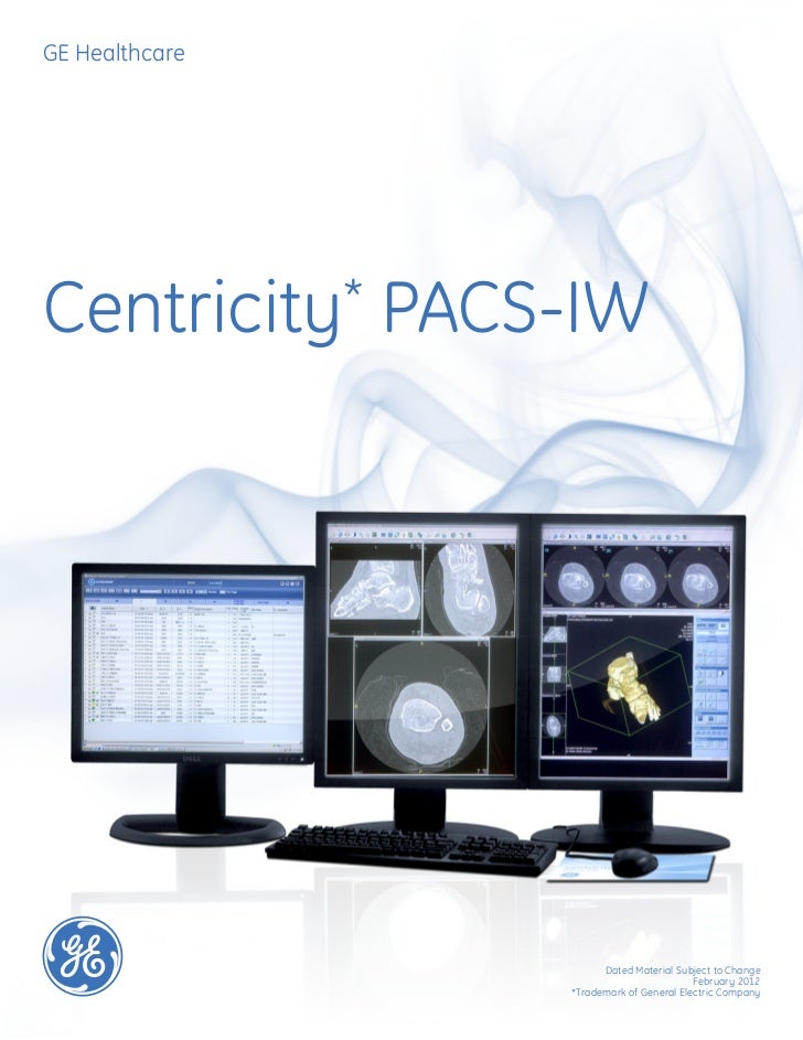
Luke’s Hospital and Health Network in Bethlehem, Pa., is a nationally recognized tertiary care facility that is committed to high-tech, high-quality patient care. That commitment extends across the enterprise. The radiology department is a center of excellence employing state-of-the-art technology. Two solutions that keep the department on the cutting edge of patient care are GE Healthcare Centricity PACS and Centricity AW Suite 2.0. These solutions also meet the second key need in radiology: streamlined workflow. Two years ago, the hospital was among the first adopters of the GE Centricity PACS and Centricity AW Suite 2.0, and since then it has real significant benefits in terms of convenience, flexibility, efficiency and workflow.
Ge Centricity Pacs Manual Meatloaf
“We use AW Suite on Centricity PACS workstations for CT and MRI reconstructions. The combination allows us to manipulate images in any plane without transferring the dataset from PACS to a freestanding AW and back again,” explains Laurie Sebastiano, MD, radiologist and clinical director of emerging technologies.
One of the primary advantages AW Suite brings is the essential AW 3D rendering tools to GE Centricity PACS, which transforms workflow in the busy department. “Many sites have only one or two standalone 3D post-processing workstations, which aren’t always in the most convenient spot,” notes Sebastiano. And workflow processes can be clunky.
Technologists, who aren’t always sure which studies require 3D manipulations, must anticipate radiologists’ needs and push the datasets to the standalone workstation. Then the radiologist needs to move to the AW and hope that the station is unoccupied. The radiologist completes the 3D manipulations, saves the new images and sends them back to PACS before returning to the PACS workflow.
“With AW Suite, it’s much more free flowing,” sums Sebastiano. The radiologist can recall images needed for 3D reconstruction from any Centricity workstation, without moving from the PC or interrupting conventional PACS workflow. There is no need to push data or send images back to PACS. Inside advice St. Luke’s radiology department has optimized its Centricity AW Suite 2.0 deployment via several key processes.
“It’s important that sites outline protocols for data use,” says Sebastiano. “It’s not particularly effective if the site sends 5 millimeter reconstructions to PACS.” On the flip side, thinner slices are useful, but tend to gobble memory and hog the archive. Luke’s addressed the situation by upgrading Centricity workstations to dual processors. In addition, although 2 gigabyte memory suffices for 3D rendering, several users were upgraded to 4 gigabyte memory for optimum performance. The result is a fast, responsive experience, says Sebastiano.
Datasets typically load in under 20 seconds, and the tools such as grab and drop react to the user with virtually no delay. The other key to system performance is vendor service and support.
“We run a lot of programs on Centricity and work closely with GE to ensure integration and optimum performance,” states Sebastiano. For example, St.
Luke’s relies on an integrated third-party voice recognition system on its PACS workstation. The combination is another workflow booster as it automatically loads demographic information into the dictation system and allows the radiologist to begin dictating immediately — focusing on the meat of radiology rather than data entry or IT tasks. Similarly, the radiology department runs Confirma’ CADstream breast MRI CAD software on its Centricity PACS desktops with CADstream images on one screen and Centricity images on the other two.

“The convenience is terrific,” says Sebastiano. “Radiologists don’t have to disrupt their workflow and walk to the CADstream server to interpret breast MRI studies.” Future growth The GE/St. Luke’s partnership has delivered significant benefits. The ability to integrate Centricity with third-party systems translates into efficient workflow processes across the radiology department. In addition, the Centricity AW Suite 2.0 solution re-invents 3D post-processing by bringing complete 3D functionality to the radiologists’ home base — the PACS workstation.
Separate trips to a standalone workstation for 3D manipulation are unnecessary, which enhance convenience and efficiency. Luke’s aims to up the ante and deploy AW RemoteAccess later this spring.
The solution crosses one of the final barriers in distributed 3D imaging. That is, it delivers AW functionality to remote Centricity portal, allowing radiologists to manipulate 3D datasets from anywhere. “The additional workflow gains will be significant for radiologists on call,” predicts Sebastiano.
Featuring a single image repository across 2D and 3D studies, Centricity Universal Viewer intuitively brings together the tools needed by radiologists, cardiologists and other clinicians to provide enterprise-wide access on a single desktop. Centricity Universal Viewer delivers:. Intelligent productivity tools.
Advanced visualization applications. An advanced mammography workflow.
Cross Enterprise Display. Advanced Cardiology tools. Access from anywhere 1. Image enable your EMR 1 Where an internet connection is available.
2 Refer to the list of supported devices and supported browsers in the technical information section of this datasheet. Smart Reading Protocols A standard feature of Centricity Universal Viewer, Smart Reading Protocols (SRP) represents a different way for radiologists to create hanging protocols and improve image setup efficiency. In contrast to traditional hanging protocol functions available with PACS software, SRP does not require the user to set up hanging protocol rules that anticipate a multitude of scenarios, exam types and procedure information. During the normal review process, radiologists hang images based on their preferences and simply “teach” the system using a one-click learning feature.
The next time the radiologist opens an exam of a similar type, SRP automatically applies the previous learnings to “hang” the image to the specified preference. Smart Reading Protocols are available in multiple languages. Adaptive streaming engine The adaptive streaming engine achieves rapid image display by adjusting for network conditions, device compute power, and image compression to provide the most efficient way to deliver images. It responds rapidly to user actions to prioritize images most likely to be displayed next by the user, and uses progressive image quality if needed to optimize the user experience. Centricity Universal Viewer provides an enhanced user experience for advanced visualization.
It offers a single source of applications and toolsets for multiple modalities and care areas, with native applications for MIP/MPR and PET-CT and enhanced solutions for vessel analysis, automated bone removal, automated registration, oncology quantification, lung and colon analysis, CT perfusion and others. The embedded advanced applications decrease the need to log in to multiple applications and retrieve images for comparison from stand-alone “mini PACS” systems, allowing radiologists and clinicians to read exams more efficiently. This also helps increase IT productivity by reducing requests for DICOM resends and image moves between systems. The embedded applications are combined with the system’s hanging protocols and the smart reading protocols to help reduce manual per-exam setup and support rapid reading. Preprocessed images are displayed on the monitor according to the radiologist’s preset specifications. The starting point for the embedded applications is the 3D Advantage package. This package provides multi-modality tools used in the radiologist’s daily work.
Key features include:. 3D visualization and processing capabilities for reading and comparing CT, MR, 3D X-ray, PET, PET/MR and PET/CT datasets. A broad portfolio of high-performance analysis tools, automating routine tasks and helping to make 3D image processing a stress-free component of your routine workflow. Improved vessel visualization by removing obstructive bony detail. The capability to align and fuse two volumetric acquisitions from either the same or different acquisition modalities, allowing you to easily compare 3D anatomical images from CT, MR with PET, and X-ray angiography for a comprehensive analysis.
Full support of PET Standard Uptake Value (SUV). Centricity Universal Viewer’s breast imaging workflow application provides access to a broad set of imaging tools, supports screening and diagnostic workflows and the display of multimodality images on the same workstation. This distinctive application allows mammographers and radiologists to rapidly access patient history and relevant priors along with the ability to read all image types available for the patient on the same workstation, helping reduce the need to maintain separate, stand-alone workstations and separate specialized systems. For organizations without dedicated mammographers, breast images can be read as part of the overall workflow.
The mammography and breast imaging workflow application features softcopy reading with customizable protocols and CAD display. Image types supported include mammography, tomosynthesis, breast MR, breast ultrasound, and Contrast Enhanced Spectral Mammography (CESM). Non-breast images available in the system can be displayed to help provide more information on the patient. Breast images are available for cardiologists to view to help enhance diagnosis and treatment recommendations. Put multi-modality images at your fingertips to help deliver a primary diagnosis faster. Centricity Universal Viewer Zero Footprint (ZFP) provides clinicians, radiologists, cardiologists and specialists enterprise-wide and community-wide access to images and reports from anywhere1 on the user’s device of choice2.
Centricity Universal Viewer ZFP provides access to direct or through connectivity with your institution’s Electronic Medical Record (EMR), HIS, RIS or physician portal applications. Centricity Universal Viewer ZFP is cleared for diagnostic use3 and helps physicians view the clinical content that is available for a patient across the longitudinal care continuum. Both DICOM data and XDS (Cross–Enterprise Document Sharing) content can be viewed in a single solution. 1 Where an internet connection is available. HIS and RIS as well as stand-alone access 2 Refer to the list of supported devices and supported browsers in the technical information section of this datasheet. 3 Centricity Universal Viewer ZFP client has been validated and cleared for diagnostic use by the US FDA on Microsoft ®Windows ® and Apple ® Mac ® products. ZFP has also received CE Mark for diagnostic use.
As regulatory clearance requirements differ by country and region, GE Healthcare must obtain clearance in countries where local specific regulatory approvals are required. Your sales representative can provide information on the status of availability in your area.
ZFP can also be used on the Apple ® iPad, ® Samsung Galaxy Note ® 10.1 and Galaxy Tab ® 4 in a review only mode and is not meant for primary diagnosis on these devices. Please refer to the product datasheet for a list of operating systems and browsers supported on these devices. Universal Viewer with Cardiology capabilities. Centricity Universal Viewer provides advanced visualization and quantification tools for Cardiology, offering a single source for coronary and vascular analysis, volume analysis, echo measurement and analysis such as 2D strain, 3D and 4D visualization as well as advanced measurement tools for CATH procedures. Advanced Cardiology Tools are powered by TomTec-Arena TM1 Additional powerful tools that are tightly connected with Centricity Universal Viewer help to enhance clinical confidence and further facilitate information flow through the department or enterprise by saving advanced analysis results as a DICOM SR (Structured Report) and a DICOM SC (Secondary Capture). With the tight connectivity to the TomTec Arena TM software, Centricity Universal Viewer provides easy and quick review of individual images and sequences supported by a variety of time saving features. Prior studies can easily be compared with current examinations including the simultaneous display of cardiac catheter and echo or NM examinations to provide additional clinical information.
1 - Provided by TomTec Imaging Systems GmbH, Edisonstrasse 6, 85716 Unterschleissheim, Germany. LEARN Radiologists and clinicians are seeking new ways to boost productivity, optimize scarce resources, and connect their workflow across the care team to help improve patient outcomes. One way to help meet these needs is to provide rapid access to images and reports when and where 1 needed through a single viewer. Learn about Centricity Universal Viewer with Imaging Desktop, a workflow solution designed to improve the radiologist reading experience by providing the right exams at the right time with access to clinical information and images that provides insights. As part of the global shift to coordinated care and collaborative care models, imaging is moving from siloed, departmental systems to assume a true, enterprise level priority. To be productive and efficient while managing cost, quality and remote/internet access, imaging departments must radically rethink their approach.

This includes customized workflows and advanced visualization. All this to help the enterprise improve clinical, financial and operational outcomes.
View this webcast on how an adaptable portfolio of technologies, insights and services can help meet the unique breast care needs of all women, especially those with elevated risks. View this webinar featuring Huron Regional Medical Center and learn what capabilities organizations should look for in a Zero Footprint viewer.
Additionally you will learn a best practice approach to adopting Universal Viewer ZFP and how a regional medical center quickly grew referrer access using the Universal Viewer ZFP. View this webinar recording to learn how you can help enhance diagnostic speed and confidence with GE Healthcare's Enterprise Imaging Solutions. This webinar focuses on Universal Viewer smart reading protocols; one of the intelligent tools to optimize productivity. Smart Reading Protocols (SRP) represent a breakthrough in the way radiologists work with Hanging Protocols (HP) to help reduce image setup inefficiencies. Learn how GE Healthcare is working with leading organizations to leverage Artificial Intelligence (AI) to shape the future of radiology.
This session examines the capabilities of Universal Viewer to make clinical insights accessible in one place for the user. Centricity™ Universal Viewer 100 edition is designed for smaller healthcare providers who have limited IT resources.
It is a turnkey web-based PACS solution designed to view, store and distribute images. All of the standard features of Centricity Universal Viewer are included, however, certain modules, such as Cross Enterprise Display and Advanced Cardiology are only available with the Enterprise edition. A sample comparison of the two offerings is depicted here. Further information is available by downloading the. The 100 edition is offered to providers that perform up to 100k exams a year in the US, or globally, to those providers who have a maximum of 6 radiologists. The 100 edition can easily scale to the enterprise edition should providers plan grow and expand. To learn more about Centricity Universal Viewer 100 edition, contact your GE Healthcare sales representative or an authorized reseller.
To find a reseller, click.